 With 41 years of experience, American Imaging is proud to provide our services to Physicians and Hospitals throughout the entire country.
Do not hesitate to contact us today to request information.
Nerve Conduction Velocity
A nerve conduction velocity test, also called a nerve conduction study, measures how quickly electrical impulses move along a nerve. It is often done at the same time as an electromyogram, in order to exclude or detect muscle disorders.
A healthy nerve conducts signals with greater speed and strength than a damaged nerve. The speed of nerve conduction is influenced by the myelin sheath—the insulating coating that surrounds the nerve.
Most neuropathies are caused by damage to the nerve’s axon rather than damage to the myelin sheath surrounding the nerve. The nerve conduction velocity test is used to distinguish between true nerve disorders (such as Charcot-Marie-Tooth disease) and conditions where muscles are affected by nerve injury (such as carpal tunnel syndrome).
This test is used to diagnose nerve damage or dysfunction and confirm a particular diagnosis. It can usually differentiate injury to the nerve fiber (axon) from injury to the myelin sheath surrounding the nerve, which is useful in diagnostic and therapeutic strategies.
During the test, flat electrodes are placed on the skin at intervals over the nerve that is being examined. A low intensity electric current is introduced to stimulate the nerves.
The velocity at which the resulting electric impulses are transmitted through the nerves is determined when images of the impulses are projected on an oscilloscope or computer screen. If a response is much slower than normal, damage to the myelin sheath is implied. If the nerve’s response to stimulation by the current is decreased but with a relatively normal speed of conduction, damage to the nerve axon is implied.
There is generally minimal discomfort with the test because the electrical stimulus is small and usually is minimally felt by the patient.
Evoked Potentials
Evoked potentials are used to measure the electrical activity in certain areas of the brain and spinal cord. Electrical activity is produced by stimulation of specific sensory nerve pathways. These tests are used in combination with other diagnostic tests to assist in the diagnosis of multiple sclerosis (MS) and other disorders.
Evoked potentials test and record how quickly and completely the nerve signals reach the brain. Evoked potentials are used because they can indicate problems along nerve pathways that are too subtle to show up during a neurologic examination or to be noticed by the person. The disruption may not even be visible on MRI exam.
General Ultrasound
Arterial
Carotid
Duplex Arterial
Transcranial Doppler
Venous
Musculoskeletal Ultrasound
American Imaging also provides musculoskeletal and soft-tissue ultrasound testing for the cervical, thoracic, and lumbar areas, along with extremities of shoulders, elbows, wrists, knees, and ankles.


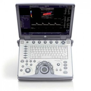
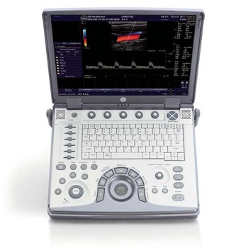
 GE LOGIQ e
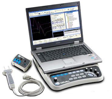
 CADWELL SIERRA WAVE


